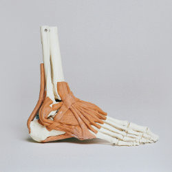
Eperon de Lenoir ou Enthésopathie (1) calcanéenne


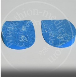
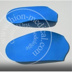
Pour Hallux valgus ou oignon du pied
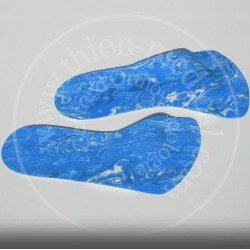
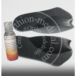
Semelles META VALGUS pour Hallux Valgus + Huile ALPHA LUX
Semelles pour soulager l'oignon du pied et huile de massage pour calmer les douleurs
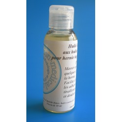
Conditionnement: 1 paire + 1 flacon de 50 ml
Adaptées aux chaussures de ville